Martensiomyces
MARTENSIOMYCES Meyer, 1959 (Bull. Soc. Mycol. France 73:189), 1 sp. (Meyer, 1957; Benjamin, 1959—ILLUS; Young, 1968a, 1968b).
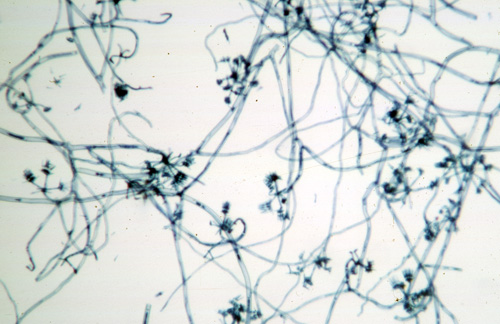
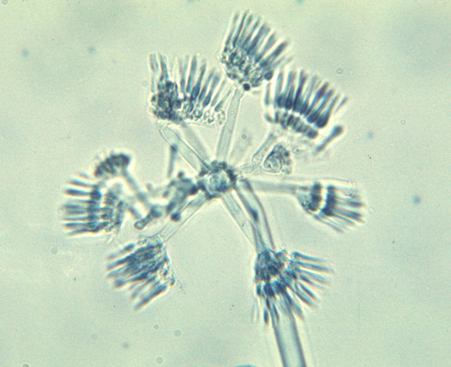
Sporangiophores septate, branched, fertile region more or less straight to curved. Sporocladia produced umbellately and successively on the vesiclate apex of a lateral branch from the fertile hyphae that grows on to produce many free, strerile apices. Sporocladium multicelled, producing pseudophialides from one surface. Merosporangia unicelled, long obclate, one produced from each pseudophialide; released in a droplet of fluid at maturity. Zygospores unknown.
Type species: M. pterosporus
Species of Martensiomyces:
M. pterosporus Meyer, 1959 (Bull. Soc. Mycol. France 73:190) (Benjamin 1959; Young 1968a, 1968b).
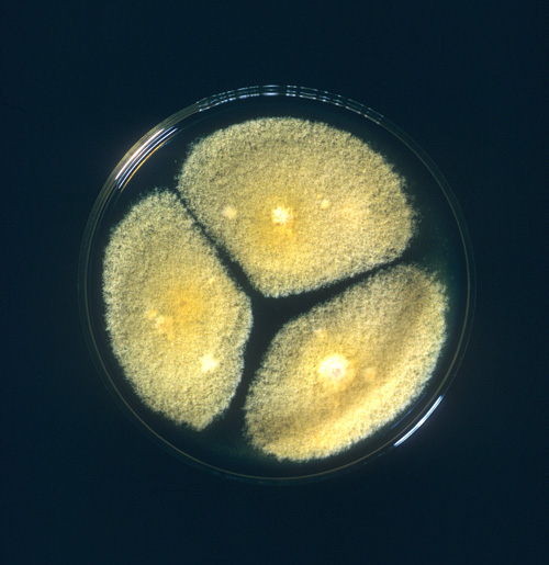
Martensiomyces forms a low growing yellow colony with the sporulating heads scattered in the aerial hyphae. Sporulation appears to be optimal on YpSs agar (Benjamin, 1959) when the fungus is incubated at 25 C. This is another taxon that was isolated from soil. It is known only from the original description (Meyer, 1957). Scanning EM photographs of M. pterosporus have been published (O’Donnell, 1979).
Bibliography
Benjamin, R.K. 1959. The merosporangiferous Mucorales. Aliso 4:321-433.
Meyer, J. 1957. Martensiomyces pterosporus nov. gen. nov. sp. nouvelle Kickxellacée isolee du sol. Bull. Soc. Mycol. France 73: 189-201.
O’Donnell, K.L. 1979. Zygomycetes in Culture. Palfrey Contributions in Botany No. 2. Department of Botany, University of Georgia, Athens, Georgia.
Young, T.W.K. 1968a. Electron microscopic study of the asexual structures in Kickxellaceae. New Phytologist 67:823-836.
Young, T.W.K. 1968b. Electron microscopic study of the asexual structures in Mucorales. Proc. Linn. Soc. London 179:1-9.
Updated Feb 26, 2007