Cokeromyces
COKEROMYCES Shanor, 1950 (In Shanor, Poitras, and Benjamin, Mycologia 42:272); 1 sp. (Shanor et al., 1950; Benny and Benjamin, 1976).
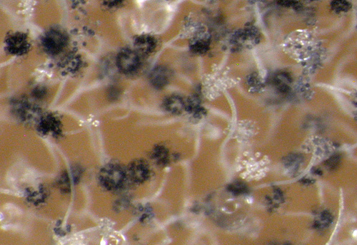
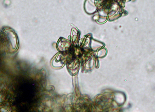
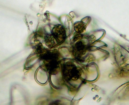
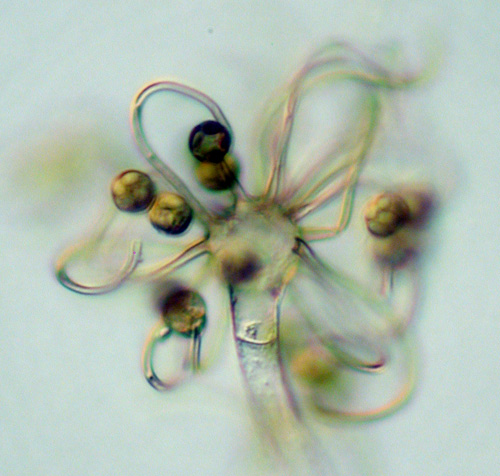
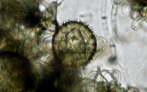
Cokeromyces produces relatively short sporangiophores that arise directly from the substrate and the apex terminates in a single vesicle that bears several recurved to twisted and contorted pedicels. Sporangia are formed at the apex of each pedicel that are globose, columellate, multispored, and they have a persistent wall. Zygospores with rough, dark walls and opposed suspensors are formed just above the surface of the substrate; the fungus in homothallic.
Type species: C. recurvatus
Species of Cokeromyces:
C. recurvatus Poitras, 1950 (in Shanor, Poitras, and Benjamin, Mycologia 42:272).
Cokeromyces recurvatus was described by Shanor et al. (1950) from Illinois and Gilman et al. (1957) isolated it from Iowa. Benny and Benjamin (1976) studied specimens of C. recurvatus from Mexico (Baja California) and the U.S.A. (Arizona, California, Florida, Illinois, Michigan, Texas); all cultures were isolated from dung except the one made in Texas (soil). Cokeromyces was redescribed and illustrated by Benny and Benjamin, 1976). The colony of C. recurvatus is low growing (less than 1 mm high) and dark brown in color. Cokeromyces is the host of choice for several mycoparasites in the Dimargaritales and many species of Piptocephalis. Some species of Syncephalis, for example S. depressa may also be grown on Cokeromyces recurvatus.
Benny et al. (1985) discussed yeast formation in C. recurvatus. Ultrastructural studies on Cokeromyces have been published by Jeffries and Young (1983a, 1983b). O’Donnell (1979) published SEM photographs of C. recurvatus.
Poitras (1957) found that zygospores and sporangia were formed when grown under 24 hr of fluorescent light but under alternating fluorescent light and dark the colony formed rings because sporangia were not produced without light. Direct sunlight inhibited sporulation and placing the cultures close to a window receiving incidental sunlight induced sporangial formation but inhibited zygospore production. Incubation temperature (Poitras, 1957) was thought to also be important for reproduction of C. recurvatus. The best growth and sporulation of C. recurvatus was obtained when grown on Emerson’s YpSs agar (Difco) and MEYE agars at 26 C (Benny and Benjamin, 1976). Growth rate was about the same at 26 C and 31 C but there was little growth at 36 C (G. Benny, unpublished data, 1971).
Cokeromyces recurvatus also is known be a rare cause of mucormycosis in humans (Axelrod et al., 1987; Alvarez et al., 1995; Munipalli et al., 1996; Tsai et al., 1997). There also have been papers published on the isolation, identification, and storage of C. recurvatus in the clinical laboratory (McGough et al., 1990; Pasarelli and McGinnis, 1992; Kemna et al., 1994).
Bibliography
Alvarez, O.A., J.A. Maples, F.O. Tio, and M. Lee. 1995. Severe diarrhea due to Cokeromyces recurvatus in a bone marrow transplant recipient. AJG 90:1350-1351
Axelrod, P., K.J. Kwon-Chung, P. Frawley, and H. Rubin. 1987. Chronic cystitis due to Cokeromyces recurvatus: a case report. Journal of Infectious Diseases 155:1062-1064.
Benny, G.L., and R.K. Benjamin. 1976. Observations on Thamnidiaceae (Mucorales). II. Chaetocladium, Cokeromyces, Mycotypha, and Phascolomyces. Aliso 8: 391-424.
Benny, G.L., P. M. Kirk, and R. A. Samson. 1985. Observations on Thamnidiaceae (Mucorales). III. Mycotyphaceae fam. nov. and a re-evaluation of Mycotypha sensu Benny & Benjamin illustrated by two new species. Mycotaxon 22: 119-148.
Jeffries, P., and T.W.K. Young. 1983a. Light and electron microscopy of vegetative hyphae, septum formation, and yeast-mould dimorphism in Cokeromyces recurvatus. Protoplasma 117:206-213.
Jeffries, P., and T.W.K. Young. 1983b. Zygospore structure in Cokeromyces recurvatus with notes on the asexual apparatus. Mycologia 75:509-517.
Kemna, M.E., R.C. Neri, R. Ali, and I.F. Salkin. 1994. Cokeromyces recurvatus, a mucoraceous zygomycete rarely isolated in clinical laboratories. J. Clin. Microbiol. 32:843-845.
McGough, D.A., A.W. Fothergill, and M.G. Rinaldi. 1990. Cokeromyces recurvatus Poitras, a distinctive zygomycete and potential pathogen: criteria for identification. Clin. Microbiol. Newsletter 12:113-117.
Munipalli, B., M.G. Rinaldi, and S.B. Greenberg. 1996. Cokeromyces recurvatus isolated from pleural and peritoneal fluid: case report. J. Clin. Microbiol. 34:2601-2603.
O’Donnell, K.L. 1979. Zygomycetes in culture. Palfrey Contributions in Botany. No. 2. Department of Botany, University of Georgia, Athens, Georgia. 257 p.
Pasarell, L., and M.R. McGinnis. 1992. Viability of fungal cultures maintained at –70C. J. Clin. Microbiol. 30:1000-1004.
Shanor, L., A.W. Poitras, and R.K. Benjamin. 1950. A new genus of the Choanephoraceae. Mycologia 42:271-278.
Tsai, T.W., L.A. Hammond, M. Rinaldi, K. Martin, F. Tio, J. Maples, C.O. Freytes, and G.D. Roodman. 1997. Cokeromyces recurvatus infection in a bone marrow transplant recipient. Bone Marrow Transplant. 19:301-302.
Updated Mar 14, 2005