Blakeslea
BLAKESLEA Thaxter, 1914 [Bot. Gaz. (Crawfordsville) 58:353]; 3 spp. (Kirk, 1984—monograph; Zheng and Chen, 1986).
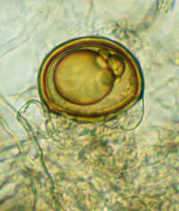
Blakeslea (Kirk 1984) produces two types of sporangia on separate sporangiophores. The large sporangia are like those of Gilbertella persicaria (see below). Few-spored sporangia are one- to eight-spored and they are borne on barrel-shaped pedicels that cover the surface of globose vesicles. The wall of the small sporangium has a suture in it and, therefore, the spores and the sporangial wall are readily separable, even when only a single spore is present. The sporangiospores are fusiform, pigmented, and striate with several long, hyaline appendages arising from the ends. The zygospores are borne on apposed (parallel) suspensors that are more or less wrapped around one another. The zygospore wall is hyaline and smooth whereas the zygosporangial wall is pigmented and striate. The zygospores form in or on the substrate.
Type species. B. trispora
Species of Blakeslea:
B. monospora B.S. Mehrotra & Baijal, 1968 (J. Elisha Mitchell Sci. Soc. 84:207).
B. sinensis Zheng & G.Q. Chen, 1986 (Acta Mycol. Sinica Suppl. 1:45).
B. trispora Thaxter, 1914 [Bot. Gaz. (Crawfordsville) 58:353].
The type species of Blakeslea, B. trispora (Thaxter, 1914), was originally isolated from a caterpillar on cowpea that was infected with Nomuraea rileyi (as Botrytis rileyi) sent to Harvard University from Gainesville, Florida; Gainesville is the type (topotype) locality for B. trispora. Good illustrations for B. trispora have been published by Thaxter (1914), Benny and O’Donnell (1978; as Choanephora trispora), and Kirk (1984). A third species, B. sinensis (Zheng and Chen, 1986) was isolated from flowers, paper, and soil in China.
Blakeslea trispora has been used to study trisporic acid biosynthesis (Sutter and Whitaker, 1981; Sutter and Jelinek, 1983). Benny and O’Donnell (1978) used light and scanning electron microscopy (SEM) to observe both types of mature sporangia. sporangiospores, and zygospore ontogeny. Mistry (1977) used SEM to study the development of zygospores in B. trispora.
Blakeslea trispora is commonly isolated from soil collected in Florida, and other parts of the southeastern United States especially in the summer. In southeast Asia (India, China) B. trispora is reported to be a pathogen of various plants. A second species, B. monospora (B.S. Mehrotra and Baijal, 1968) was described from India. A second isolate of B. monospora was reported from Taiwan (Ho and Chang, 2003).
Bibliography
Benny, G.L., and K.L. O’Donnell. 1978. Choanephora trispora, pp. 135-136. In: M.S. Fuller (ed.). Lower fungi in the laboratory. Palfrey Contributions in Botany. No. 1. Department of Botany, University of Georgia, Athens, Georgia.
Ho, H.-M., and L.-L. Chang. 2003. Notes on Zygomycetes of Taiwan (III). Two Blakeslea species new to Taiwan. Taiwania 48:232-238.
Kirk, P.M. 1984. A monograph of the Choanephoraceae. Mycol. Paper 152:1-61.
Mehrotra, B.S., and U. Baijal. 1968. Is Blakeslea a valid genus? J. Elisha Mitchell Sci. Soc. 84:207-210.
Mikawa, T. 1979. A taxonomic study on Japanese sporangiferous Mucorales (6). J. Japan. Bot. 54:289-300.
Mistry, A. 1977. Zygosporogensis in Blakeslea trispora (Mucorales). Microbios 20:73-79.
Sutter, R.P., and B.G. Jelinek. 1983. Trisporic acid biosynthesis in cultures of Blakeslea trispora with (+) and (-) mycelia separated by a filter. Exper. Mycol. 188-191.
Sutter, R.P., and J.P. Whitaker. 1981. Sex pheromone metabolism in Blakeslea trispora. Naturwissenschaften 68:147.
Thaxter, R. 1914. New or peculiar Zygomycetes. 3: Blakeslea, Dissophora and Haplosporangium, nova genera. Bot. Gaz. (Crawfordsville) 58:353-366.
Zheng, R.-y., and G.-q. Chen. 1986. Blakeslea sinensis sp. nov., a further proof for retaining the genus Blakeslea. Acta Mycol. Sinica Suppl. 1:40-55.
Updated Mar 14, 2005