Phascolomyces
PHASCOLOMYCES Boedijn, 1959 (Sydowia 12:349); 1 sp. (Benny and Benjamin, 1976—monograph).
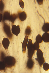
Phascolomyces forms simple or branched sporangiophores that form lateral or terminal fertile vesicles bearing many unispored sporangia on long, thin, more less straight pedicels. Relatively large pigmented chlamydospores are produced in the substrate. Zygospores are unknown (Benny and Benjamin, 1976).
Type species: P. articulosus
Species of Phascolomyces:
P. articulosus Boedijn, 1959 (Sydowia 12:349) ex Benny & R.K. Benjamin, 1976 (Aliso 8:417).
Phascolomyces articulosus can be confused with Cunninghamella spp. when first observed on the natural substrate (dung). Phascolomyces was included in the Cunninghamellaceae (Mucorales) by Boedijn (1958), Mil’ko and Beljakova (1967), Hesseltine and Ellis (1973), and Mil’ko (1974). Benny and Benjamin (1976) transferred P. articulosus to the Thamnidiaceae and validated the taxon.
The spore wall of P. articulosus is bilaminate and it is verrucosely ornamented. Phascolomyces is unusual in producing a trilaminate, sporangial wall (Jeffries and Young, 1978b). Phascolomyces, of all of the Mucorales tested, is not parasitized by Piptocephalis unispora (Jeffries and Young, 1978a). Jeffries and Young (1978a) observed that appressoria, penetration pegs, and haustoria are formed by P. articulosus but the infection by P. unispora did not procede beyond that stage. Phascolomyces articulosus produces cysteine proteinase whereas other Mucorales produce serine proteinase. Parasitism of P. articulosus by Piptocephalis is suppressed because chitin proteinase is active at the site of infection (Balasubramanian and Manocha, 1986) but other Mucorales are parasitized because of the suppression of chitin deposition.
Bibliography
Balasubramanian, R., and M. S. Manocha. 1986. Cysteine proteinase of Phascolomyces articulosus: purification and properties. Can. J. Bot. 64:2441-2445.
Benny, G.L., and R.K. Benjamin. 1976. Observations on Thamnidiaceae (Mucorales). II. Chaetocladium, Cokeromyces, Mycotypha, and Phascolomyces. Aliso 8: 391-424.
Boedijn, K. B. 1958 [1959]. Notes on the Mucorales of Indonesia. Sydowia 12: 321-362.
Hesseltine, C.W., and J.J. Ellis. 1973. Mucorales, pp. 187-217, In G. C. Ainsworth, F. K. Sparrow, and A. S. Sussman (Eds.). The Fungi. Vol. 4b. Academic Press, New York. 504 p
Jeffries, P., and T.W.K. Young. 1978a. Mycoparasitism by Piptocephalis unispora (Mucorales): host range and reaction with Phascolomyces articulosis. Can. J. Bot. 56:2449-2459.
Jeffries, P., and T.W.K. Young. 1978b. Ultrastructure of the dormant and germinating sporangiospore of Phascolomyces articulosus. Can. J. Bot. 56:747-753.
Mil’ko, A.A., and L.A. Beljakova. 1967. Rod Cunninghamella Matruchot I taxonomie Cunninghamellaceae [Genus Cunninghamella Matruchot and taxonomy of the Cunninghamellaceae]. Mikrobologiya 36:684-690 [English translation available in Microbiology 36 (4):573-578, 1967].
Mil’ko, A.A. 1974. Opredeltiel’ mukoral’nykh gribov Key to the identification of Mucorales. ‘Naukova Dumka’ , Kiev, Ukraine. 303 p.
Updated Mar 13, 2005